Computed tomography (CT)
Computed tomography is a modern imaging method that enables a detailed insight into the human body.
An X-ray tube rotates around the patient, emitting at the same time a fine bunch of X-rays. Opposite to the tube, a system of detectors rotates around the patient. This helps to take pictures of the slices of the body organs from nearly a thousand of directions.
This sums up to hundred thousands of measured values that will be further processed by a computer. The result will be non-overlapping cross-sectional images of the examined region of the body. The measurement period for each body region is just a few seconds.
The examination is painless and uncomplicated for the patient.
The special advantage of CT over the conventional X-ray is a better depiction of the soft tissues, i.e. internal organs.
It is possible to examine large parts of the body in a very short time. The special recording technique helps to avoid the overlapping of the organs that appear in an X-ray image (summation image) and to make a more accurate diagnosis. Through post-processing of the measured data it is possible to produce three-dimensional images of the examined area. Furthermore, by means of density measurements one can determine whether e.g. what is seen is an adipose tissue, a fluid or a firm tissue structure.
Nowadays, computed tomography has a very wide range of application. With examinations of the lungs, many conditions of the abdominal cavity and the quick examination of the head (e.g.), there is no other method that can provide decisive information so quickly and precisely. Especially in case of accidents and strokes, computed tomography is indispensable.
Information on examinations of particular body parts:
CT thorax
CT thorax
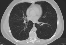
By means of computed tomography of the thoracic organs it is possible to detect early tumourous changes in the lung tissue that are still well curable. Even inflammations that are still invisible on a conventional X-ray can be clearly identified by the CT.
Another important aspect is chronic lung diseases that can be diagnosed reliably enough by means of CT only, which is in turn very important for choosing a therapeutic method.
This method can identify even occupational lung diseases (e.g. asbestosis). The bony structures of the thoracic skeleton can also be well depicted by means of special CT technique.
CT head
CT head
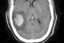
Computed tomography of the skull and brain is an important method in case of a suspected stroke and after head injuries, due to the fact that it can quickly and reliably exclude eventual brain haemorrhage, which is in turn very important for further therapy. CT can also be very helpful in case of bony changes in the skull and also for identifying fractures of the skull, skull base and facial skull.
Prior to operations of paranasal sinuses or middle ear, it is usually required to perform a CT for the exact planning of the procedure.
CT neck
CT neck
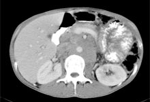
Indications: suspicion of a tumour or lymph node diseases
CT can be described as a cross-sectional X-ray of the thoracic organs. It is necessary to make a series of images.
Lying on a movable table, you will slide through the CT ring. Do not be afraid, it is not dangerous for you at all. By means of an X-ray tube and diaphragms, a thin X-ray beam is produced (fan beam). It penetrates the part of the body to be examined and is then absorbed by different structures inside the body (e.g. skin, fat, muscles, organs, bones) in varying degrees.
Exactly opposite the X-ray tube there are a large number of sensors (detectors) that receive the weakened signal, process it electronically and send to a computer for evaluation.
Directly thereafter, the X-ray tube together with the opposite-lying detectors continues slightly rotating around the patient. The described process is repeated. In this way, different perspectives of the same layer are recorded and processed into a graphic image on a computer. This image can be seen and evaluated on a monitor or an X-ray film.
CT abdomen
CT abdomen
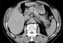
Computed tomography of the whole abdominal cavity is often applied in follow-up examinations after tumour diseases to exclude metastases in the organs or lymph nodes. Furthermore, it can be helpful in analysing liver lesions or also chronic pancreatitis that is often accompanied by fine calcifications.
Computed tomography is ideally suited to identify calcified gallstones, which makes it possible to make a decision for or against eventual medicamentous dissolution.
CT is also well suited for detecting calcified stones in the kidneys or draining urinary tracts. Bony changes in the pelvic skeleton represent another important area of application, due to the fact that they occur in case of malignant diseases.
CT spine
CT spine
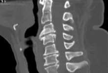
While examining the segments of the spinal column, computed tomography can give additional information about bony changes that e.g. cannot be explicitly clarified with the help of MRI. Even the smallest parts of the spinal region can be examined in order to exclude a herniated disc.
If large parts of the spinal column should be examined, MRI is considered to be the method of choice today, due to the fact that it can better depict soft tissues and does not employ X-ray radiation. Furthermore, CT is often applied in pain therapy for treating spinal disorders (see section "Therapy").
CT bone fracture
CT bone fracture
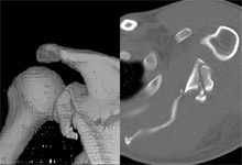
Especially after complicated bone fractures, computed tomography with its very precise cross-sectional images processed by a computer can provide a three-dimensional image of bone segments. That gives the surgeon a significantly better picture of the condition of the fracture, which can also decisively influence the therapy.
CT of Jaws/Teeth
CT of Jaws/Teeth
Thanks to renewed technical upgrading (SAFIRE Iterative Reconstruction), this computerised tomography unit now allows us to perform examinations of the jaw using dose values lower than those for known DVT devices.
The jaw examination in particular avoids unnecessary radiation exposure to the ocular lens, thus serving to avoid a cataract. The resolution limit of the device is in the area of approximately 0.33 millimetres.
Application examples:
- Unidentified pain
- Impacted wisdom teeth (prior to extraction)
- Preparation for an implant (for prevention of nerve damage)


